Molecular Inheritance & Gene Expression
A cell contains the nucleus, nucleus contains chromosomes which bear genes. Genes carry the hereditary information. A zygote has the information for development and differentiation in its genes. Cells of an individual have the genes for maintaining its structure and function.
But what are these genes and how do they function?
Genes are made of segments of the chemical DNA. This lesson deals with the study of DNA. This lesson deals with the study of DNA as the genetic material, its structure and functioning at the molecular level.
The British biochemist and physician Archibald Garrod had mentioned in his book “inborn errors of metabolism” that there are inherited genetic disorders such as phenylketonuria which are caused by of the absence of particular enzymes. Beadle and Tatum working with the mutants of the fungus Neurospora showed that the absence, of a gene in the mutant leads to absence of the enzyme in the mutant stopping a metabolic pathway (chain of biochemical reactions, midway).
Thus, was proposed that one gene was responsible for the production of one enzyme and this was called the one gene one enzyme hypothesis. Later, it was found that an enzyme may be made of more than one polypeptide and one gene-controlled production of one polypeptide.
Discovery of DNA as the Genetic (Hereditary) Material
That genes, located on chromosomes, are the hereditary material was known to scientists in the early twentieth century. That genes are segments of DNA became evident from the work of Griffith on bacterial transformation.
The bacterial Streptococcus pneumoniac when grown in the lab form smooth colonies and when injected into mice kill them. A mutant of this bacterium forms rough colonies and is harmless to mice. In 1928, Frederick Griffith found that if the smooth walled virulent form of Streptococcus is killed and mixed with the harmless rough form of. Streptococcus the letter becomes virulent (killer). This change (or transformation) of the bacteria from harmless to virulent it termed bacterial transformation.
In 1944, Avery, Mcleod and McCarty extracted the DNA from the virulent smooth steptococcus and mixed it with the non-rough variety become virulent and had a smooth coat. This did not happen when DNA of the virulent and had a smooth coat. This did not happen when DNA of the virulent was digested with the enzyme DNA and then mixed. Thus, it became clear that DNA was the transforming principle.
Later Hershey and Chase in 1952 used T2 bacteriophage (bacteria infecting virus) for their experiments. They labeled the protein coat of the virus with radioactive isotope of sulphur S35. When the virus was introduced into the bacteria, no lable (radioactivity) was found inside the bacteria as the viral coat was left outside. When they labelled viral DNA with P32 (radioactive) phosphorus, radioactivity was found inside the bacteria. It became clear that new generations of the virus were reproduced inside bacteria because of viral DNA.
These experiments confirmed that DNA is the genetic material and genes are made of Deoxyribonucleic acid or DNA.
Structure of DNA
Chemical nature of DNA
DNA is a polynucleotide, a macromolecule (macro = large) made of units called nucleotides.
Each nucleotide consists of three subunits.
(i) a pentose (5 carbon) sugar called deoxyribose
(ii) 4 nitrogenous bases Adenine (A), Guarme (G) (purine bases) and Thymine (T) and Cytosine (C) (pyrimidine bases) (Base + Sugar = Nucleoside) (Base + Sugar + Phosphate = Nucleotide) a base and a sugar combine to form a nucleoside, while it becomes a nucleotide when a phosphate group gets attached.
Base + sugar = nucleoside Heredity
Base + sugar + Phosphate = nucleotide
So, there are four nucleotides in DNA formed of sugar and nitrogenous base and phosphate.
Chargaff’s rule
The four nucleotides are not present in equal amounts in a DNA molecule. But the amount in a DNA molecule. But the amount of purines (A + G) and that of pyrimidines (T + C) is always equal. In other words, A=T and G=C. This is called Chargaff’s rule. Physical structure of DNA the DNA double helix
A DNA molecule is three dimensional and made of two strands helically coiled around each other. Franklin and Wilkins first showed through X-ray diffraction studies of DNA that is a double helix. In 1953, James Watson and Francis Crick were awarded the Nobel Prize for working out the structure of DNA.
According to Watson and Cricks model
- DNA molecule is a double helix consisting of two strands of DNA.
- The arrangement of the two strands in antiparallel, which means that the sequence of nucleotides goes up in 5′ to 3′ direction in one strand and other strand comes down in 3′ to 5′ direction. (3′ and 5′ refer to the carbon atom to which the phosphate group is attached)
- The bases of the two strands are linked by hydrogen bonds.
- Base pairing is very specific as per Chargaff’s rule. An Adenine, a Purine base always pairs with thymine, a pyridine base. The purine base Guanine pairs with the pyrimidine, Cytosine. These pairs of bases are called complementary bases.
There are two hydrogen bonds between A and T and three hydrogen bonds between G and C. In the DNA helix, a complete helical turn occurs after 3.4 nm (or 34A). This complete turn encloses 10 base pairs, each base pair lies 0.34 nm (3.4 A) apart the diameter of the double helical DNA molecule is 2.0 nm.
Watson and Crick model explains well how the two strands of a DNA molecule may separate at replication and transcription and then rewind. The hereditary material must be capsule of (i) Replication (ii) Storage of information (iii) transmitting of information (iv) expression of information (v) regulation of gene expression.
Eukaryotic Chromosome
In the bacteria (prokaryotes), only one double stranded DNA molecule constitutes the chromosome. Eukaryotes have many chromosomes and also many genes. One chromosome, however, is made up of one long double stranded DNA molecule. So how does this long molecule get accommodated in the chromosome seen as small microscopic entities during cell division.
RNA or RIBONUCLEIC ACID
Apart from DNA, RNA is the other important nucleic acid present inside the cell. Table 21.1 difference between DNA and RNA
- Double stranded molecule
- Contains deoxyribose sugar
- DNA can duplicate on its own.
Function of various type of RNA
mRNA or messenger RNA
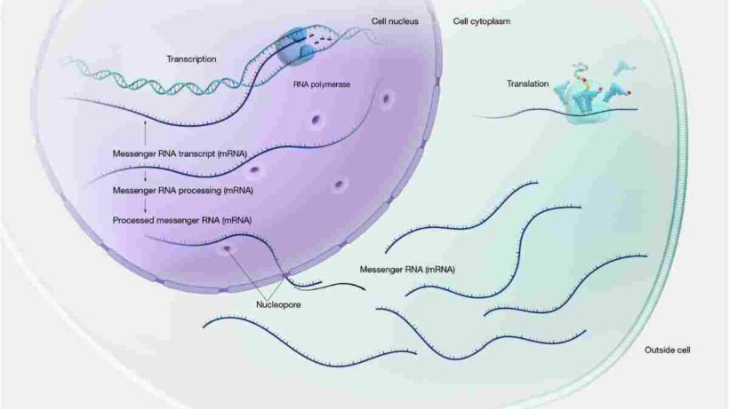
mRNA or messenger RNA is transcribed in the nucleus to carry the information for protein to be synthesized in the cytoplasm.
mRNA is transcribed as a strand of complementary bases of one of the DNA strands and carries the information for the synthesis of a particular protein or polypeptide.
tRNA or transfer RNA
tRNA or transfer RNA, also called soluble RNA has a clover leaf structure with loops. One loop recognise the ribosome, the top loop has an ‘anticodon’ to recognise the codon (triplet nucleotide sequence coding for an amino acids) on mRNA. tRNA “transfer” the amino acids at their respective positions during synthesis of protein.
There are many t-RNAs which differ in their anticodon. Each tRNA is specific for an amino acid and can carry that amino acid to the ribosome during protein synthesis.
tRNA contains unusual bases like inosine, dihyrouridine etc.
- Single stranded molecule
- contains ribose sugar. Thymine is replaced by uracil 5. RNA is synthesized on a DNA template rRNA or ribosomal RNA
rRNA is a component of ribosome which are ribonucleoprotein particles containing RNA and proteins.
rRNA have a role in protein synthesis
DNA duplicates itself exactly for passing on genetic information to the next generation of cells. Replication may thus be defined as a mechanism for transmission of genetic information generation after generation.
Mechanism of replication
Replication occurs through the following steps
- Unwinding of DNA double helix
- Synthesis of the primer. Primer is a short RNA molecule of about 5 to 10 bases.
- Synthesis of new DNA strand.
The opened strands of DNA form the template. New strands complementary to template are synthesised. At the replication fork a new DNA strand begins to synthesis, attaching itself to the primer, in the presence of the enzyme DNA polymerase. It begins synthesis from its 5′ end and contains synthesis complementary to one of the unwound parental DNA strand. The new strand of DNA continues to be synthesized to uninterrupted and is termed leading strand.
Synthesis of the other new DNA strand DNA synthesis always takes place along 5′ to 3′ direction. Therefore, the other new DNA strand gets synthesised in the direction opposite to the leading strand. This new strand, called lagging strand builds up in small piece as shown in the figure, in the presence of enzyme DNA polymerase. Thus the, synthesis of the lagging strand is discontinuous. The new piece of DNA are termed Okazaki fragments. In the presence of the enzyme ligase and the energy source ATP the Okazaki pieces get joined together to form a DNA strand
- DNA replication is remarkably accurate so that the parental DNA molecule gets an exact duplicate copy made any mistake gets chipped and repaired. This is term proof reading. At the end of results.
- DNA replication two identical DNA molecules are formed which are identical to the parent molecule.
- DNA replication is thus semi discontinuous that is one strand of the new DNA molecule builds up continuously and the other in pieces.
- DNA replication is semiconservative, since in the two new molecules formed, since parental strand is conserved and the other strand is newly synthesised experimentally proven by Meselson and Stahl.
We would love to hear your suggestions or feedback on Molecular Inheritance & Gene Expression. Feel free to share your thoughts with us!
